White Spots Multiple Sclerosis Normal Vs Ms Brain Mri Images

Imaging In Multiple Sclerosis Journal Of Neurology Neurosurgery
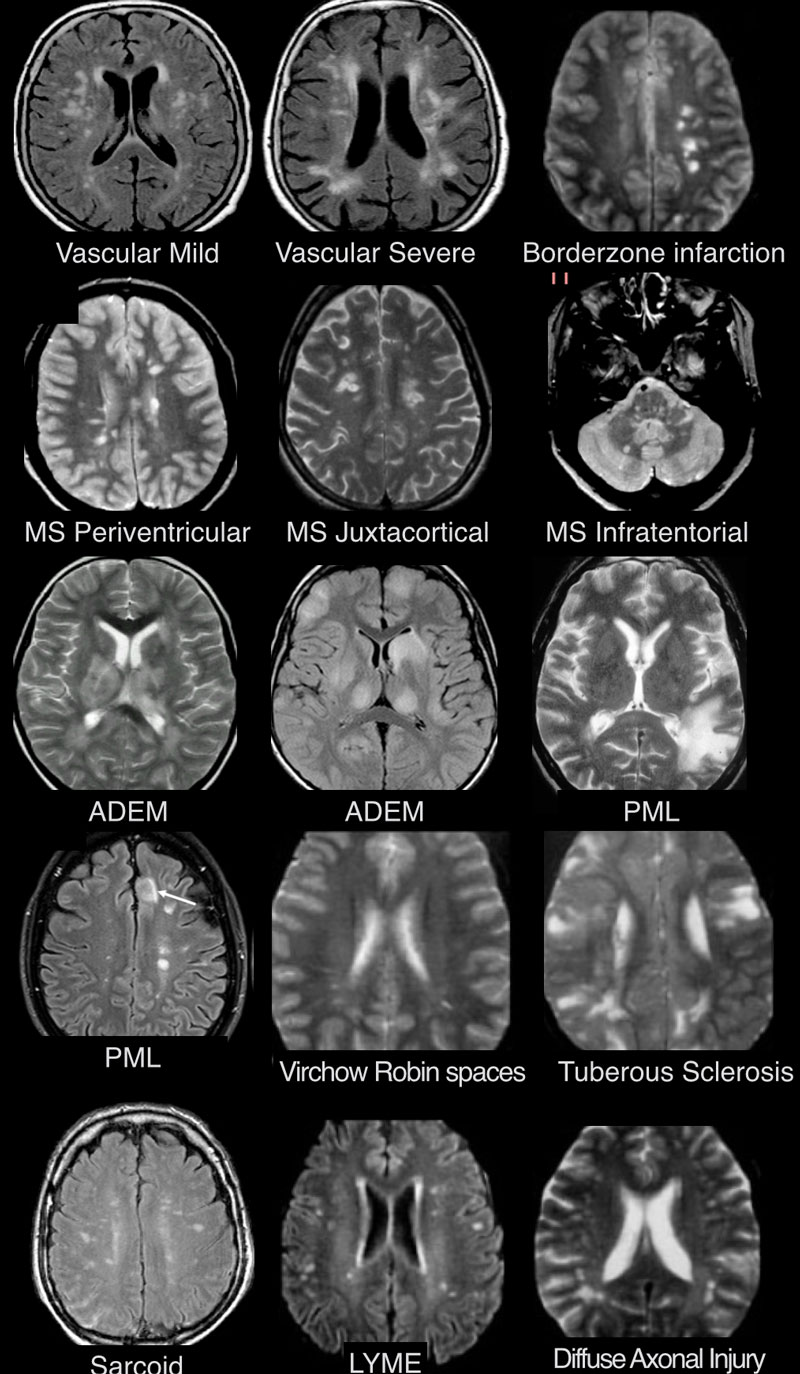
The Radiology Assistant Diagnosis And Differential Diagnosis
:max_bytes(150000):strip_icc()/what-are-these-spots-on-my-mri-2488902-5c5db0fa46e0fb0001ca86cb.png)
Spots On An Mri White Matter Hyperintensities
Https Encrypted Tbn0 Gstatic Com Images Q Tbn 3aand9gcr1ryuhls8ao8r6aab6o8r32bwv7kljtuinbg Usqp Cau

Differentiating Multiple Sclerosis Mimics On Mri Neurology Advisor
More Than Meets The Eye Multiple Sclerosis Discovery Forum

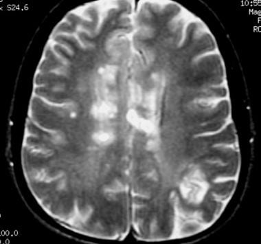
Lesions show up as white or dark spots.
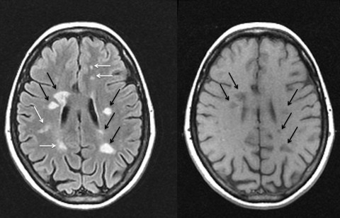
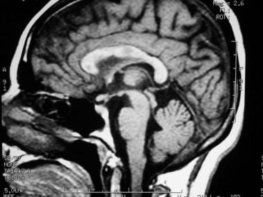
White spots multiple sclerosis normal vs ms brain mri images. Multiple small pericallosal white matter intensities characteristic of ms. At that time saggittal t2 flair imaging should be considered unfortunately my insurance wouldn t cover the mri with contrast. I was wondering if anyone else has had experience with having both chronic migraine and ms or possibly of receiving a diagnosis of migraine before a diagnosis of ms. On scanning other parts of the brain the more characteristic images of ms are seen.
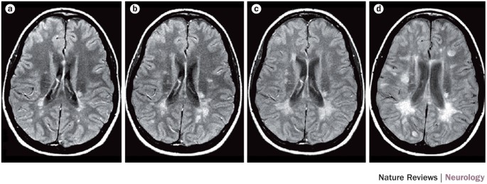
Sclerosis means scarring and people with ms develop multiple areas of scar tissue in. Ms is a chronic disease that damages the nerves in the spinal cord and brain as well as the optic nerves. Though the vast majority of ms patients have abnormalities on brain mri an estimated 5 of patients have normal imaging. Diffusion tensor imaging dti can reveal microstructural or non overt brain tissue damage and quantify pathological processes.
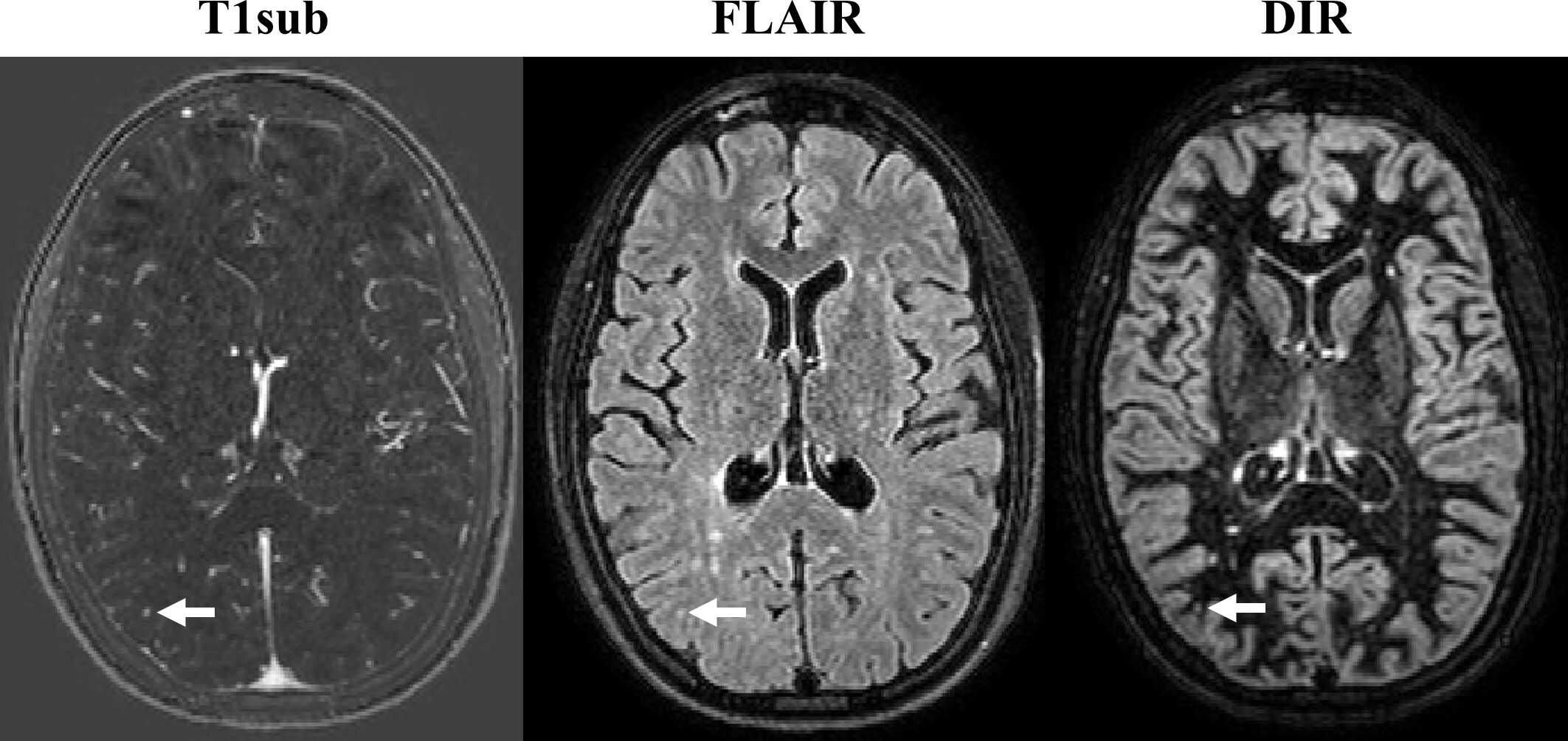
For example many disorders show up as spots on a brain mri just like ms. Ms related lesions appear on mri images as either bright or dark spots depending on the type. The afferent visual pathway represents the most frequently affected white matter pathway in multiple sclerosis ms and neuromyelitis optica spectrum disorders nmosd. Methods 67 people with relapse onset ms and 30 healthy controls were included in the study.
If you have symptoms of ms your doctor may order an mri scan of your brain and spinal cord the images produced allow doctors to see lesions in your cns. 1 alternatively there may be white matter lesions that might be seen. There are several causes of white spots on a brain mri including small strokes migraines multiple sclerosis ms lupus b12 deficiency a brain tumor such as lymphoma or an infection such as lyme disease or hiv. Mri scans can detect damage in the central nervous system which comprises the brain and spinal cord.
Volumetric t1 images and high resolution 1 mm 3 magnetisation transfer ratio mtr images were acquired and segmented into 12 bands between the inner and outer surfaces of the brain the first and last bands were discarded to limit partial volume effects with cerebrospinal fluid.
Non Contrast Mri Is Effective In Monitoring Multiple Sclerosis

Imaging In Multiple Sclerosis Journal Of Neurology Neurosurgery
Brain Imaging In Multiple Sclerosis Practice Essentials Computed
Mri Brain White Spots Cause Mri Scan Images Mri Brain Mri
Brain Imaging In Multiple Sclerosis Practice Essentials Computed

The Multiple Sclerosis Lesion Checklist Practical Neurology

Differential Diagnosis Of Multiple Sclerosis And Other

Lxdznxjugwdklm

Mri Uses In Ms Youtube

Proton Density Weighted Images From Two Subjects With Multiple

Brain Imaging In Multiple Sclerosis Practice Essentials Computed
Magnetic Resonance Imaging Is The Method Of Choice For Imaging Ms
Magnims Consensus Guidelines On The Use Of Mri In Multiple
