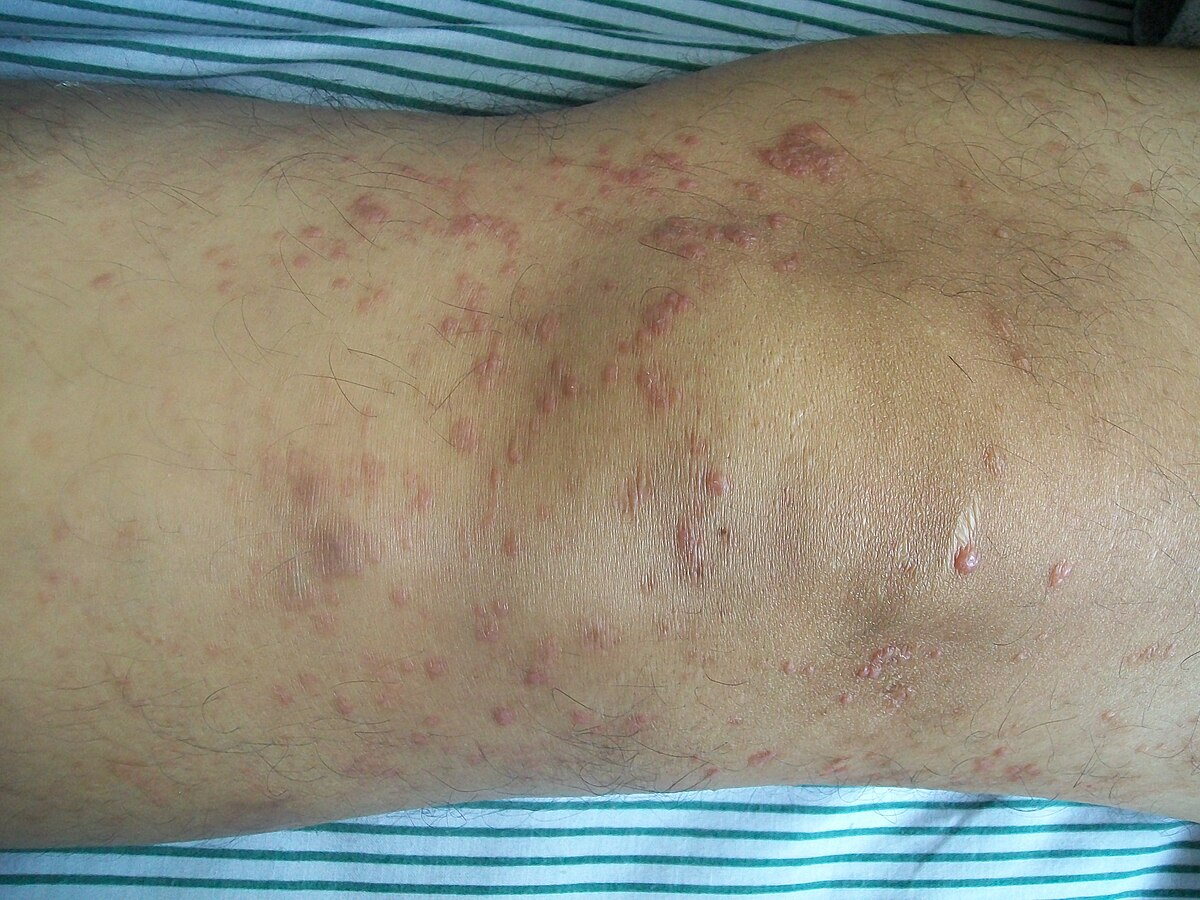
Multiple Sclerosis Lymphoma Skin Lesions

Hodgkin s lymphoma and multiple sclerosis have more than the immune system in common sharing a genetic risk factor and connection to the epstein barr virus.
Multiple sclerosis lymphoma skin lesions. Previous reports of cutaneous neoplastic lesions secondary to fingolimod treatment among multiple sclerosis patients. The patient was referred for a dermatology consultation and was scheduled for more detailed tests and a skin biopsy. The characteristic features of primary cutaneous alcl include the appearance of solitary or multiple raised red skin lesions that do not go away have a tendency to ulcerate and may itch. 298 300 cutaneous t cell lymphoma typically appears as brownish red patches with fine scale and delicate wrinkling on the skin of the trunk typically within.
It appears as skin lesions that are red to purplish large pimples plaques raised or lowered flat lesions or nodules bumps on the arms or upper body. Only about 10 percent of the time does primary cutaneous alcl extend beyond the skin to lymph nodes or organs. Skin pathology reported a diffuse infiltration of lymphoid cells in dermis and subcutis composed of cells with enlarged and prominent oval nuclei and 1 2 mitotic figures per each hpf fig. Primary cutaneous follicle center lymphoma.
Its diagnosis depends on patients history and clinical findings consisting of oral aphthae recurring at least 3 times a year plus any two findings out of genital ulcers or scars skin lesions such as erythema nodosum pseudofolliculitis or papulopustular lesions uveitis and positive pathergy test are required for the diagnosis. It accounts for most of all cases of cutaneous t cell lymphomas. This is the most common b cell lymphoma of the skin. An advanced form of mycosis fungoides is referred to as sezary syndrome.
Mycosis fungoides mf is the other common form of cutaneous lymphoma characterized by red scaly patches or lesions on the skin. Objective reporting a case of cutaneous large cell lymphoma in a multiple sclerosis patient during fingolimod treatment. There may be only a single lesion but there can sometimes be a few. Cutaneous t cell lymphomas mycosis fungoides affecting the breast or presenting initially as breast lesions are found in multiple case reports in the available literature.
It tends to grow slowly.

