Epithelioid Mesothelioma Histology
Pathology Outlines Epithelioid Mesothelioma
Pathology Outlines Mesothelioma Epithelioid
Pathology Outlines Peritoneal Malignant Mesothelioma

Mesothelioma Histology A Study Of Mesothelioma Cells
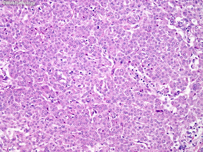
Webpathology Com A Collection Of Surgical Pathology Images
Webpathology Com A Collection Of Surgical Pathology Images
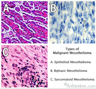
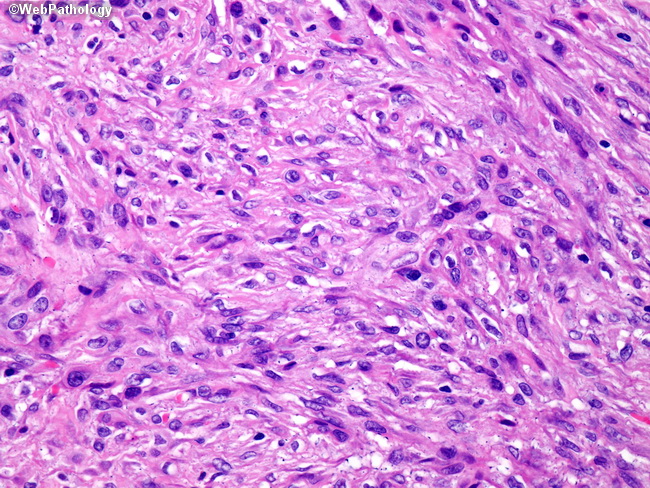
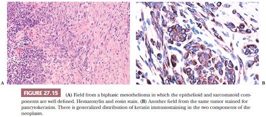
The three major mesothelioma cell types are epithelioid sarcomatoid and biphasic.
Epithelioid mesothelioma histology. Epithelial cells can develop in the lining of the lungs abdomen or heart. Epithelioid mesothelioma histology histologically epithelial mesothelioma cells have polygonal ovoid or cuboidal cell shape. Epithelioid mesothelioma cells have some notable characteristics. Mesothelioma histology or mesothelioma histopathology is the study of tissue for the presence of mesothelioma.
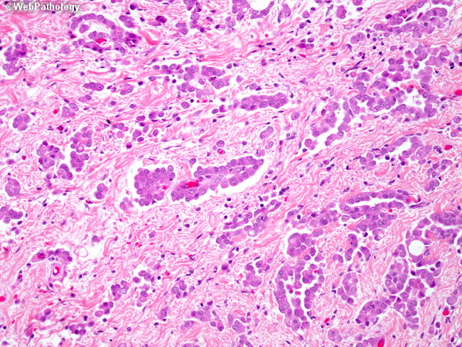
Mutations in mesothelial cells caused by asbestos exposure create these variations. Epithelioid mesothelioma is the most common mesothelioma cell type accounting for 50 to 70 of cases. Upon entering the body these asbestos fibers can lodge into cells along the mesothelium which lines the lungs abdomen and heart. This process is part of mesothelioma pathology which involves examining either tissue or fluid to determine if this cancer exists in the body.
This form of mesothelioma is comprised of cells which resemble the normal mesothelial cells in that they are arranged in a trabecular fashion. Treatment and prognosis are affected by a patient s mesothelioma cell type. Mesothelioma histology is the study of the function and structure of anatomy including tissues and cells. Epithelioid mesothelioma is caused by asbestos exposure and is the most common form of the disease accounting for 70 80 of diagnoses.
Consequently there are many diseases to be differentiated when the diagnosis of mesothelioma is based on histological analyses. The epithelial mesothelioma cell type makes up more than 50 of all cases. Another subset of this field called histopathology looks specifically at the microstructure of diseased cells and tissues which is what a pathologist will focus on when studying a tissue sample to make a diagnosis. It can take many forms.
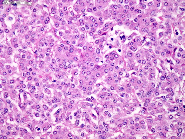
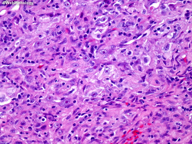
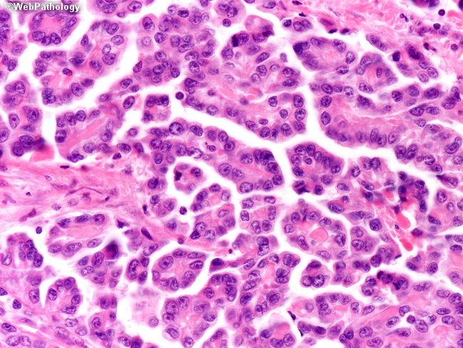
Ayesha azam for case of the week 476 ck5 6. Papillary fragments and cohesive cell clusters moderate amount of cytoplasm round to ovoid nuclei prominent nuclei. Mesothelioma can be categorized histologically as epithelioid type sarcomatoid type biphasic type desmoplastic type among others. They are square shaped cells and have visible nuclei plural for nucleus the center of the cell which carries genetic material.
The medical term histology refers to the microscopic study of tissue. Symptoms include shortness of breath and weight loss. Epithelial cells are prevalent in the human body but can be irritated by asbestos. Asbestos exposure can mutate epithelial cells which then become cancerous.
Epithelioid cell type this form of mesothelioma is the result of healthy epithelial cells in your body mutating into cancerous cells.
Webpathology Com A Collection Of Surgical Pathology Images
Webpathology Com A Collection Of Surgical Pathology Images
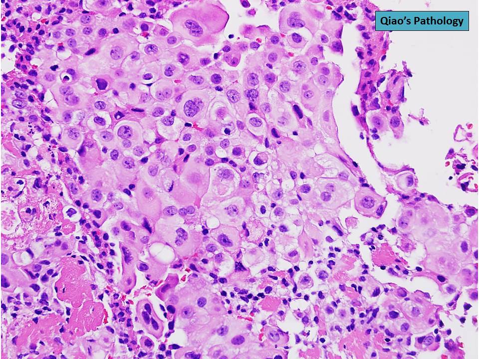
Qiao S Pathology Pleural Malignant Mesothelioma Epitheli Flickr

Primary Peritoneal Epithelioid Mesothelioma Of Clear Cell Type

Pathology Of Mesothelioma Microscopic Features Dr Sampurna Roy Md
Webpathology Com A Collection Of Surgical Pathology Images

2016 Evening Specialty Conference Gynecologic Pathology

Nuclear Grade And Necrosis Predict Prognosis In Malignant
Fig S2 Pathology Of Peritoneal Mesothelioma Microphotographs Of
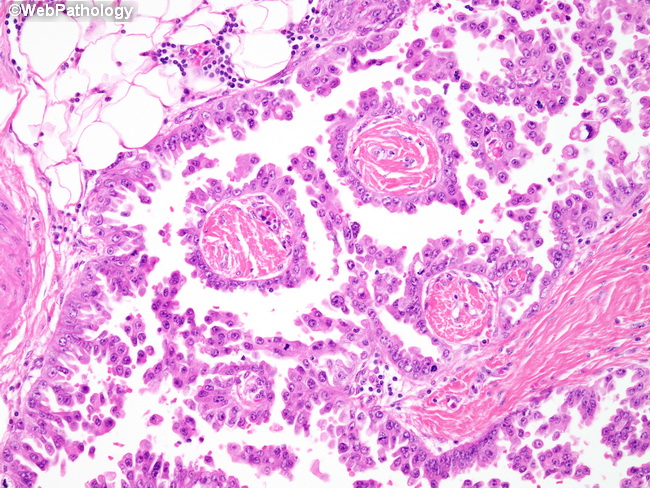
Webpathology Com A Collection Of Surgical Pathology Images
Pleura Basicmedical Key
Webpathology Com A Collection Of Surgical Pathology Images
Webpathology Com A Collection Of Surgical Pathology Images