Abnormal Ovarian Cancer Ultrasound Images
Diagnostics Free Full Text Ultrasound Monitoring Of Extant
Detection And Diagnosis Ovarian Cancer Symptoms And Diagnosis

Pelvic Ultrasound In Houston For Ovarian Cancer Screening

Full Text Transvaginal Ultrasonography In Ovarian Cancer

A Better Way To Assess Ovarian Cancer Risk Ncal Research Spotlight
The Radiology Assistant Common Ovarian Cystic Lesions
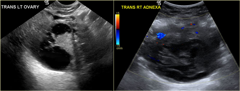
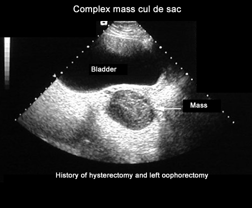
The cystic neoplasmmeasures 14 x 7 cms.
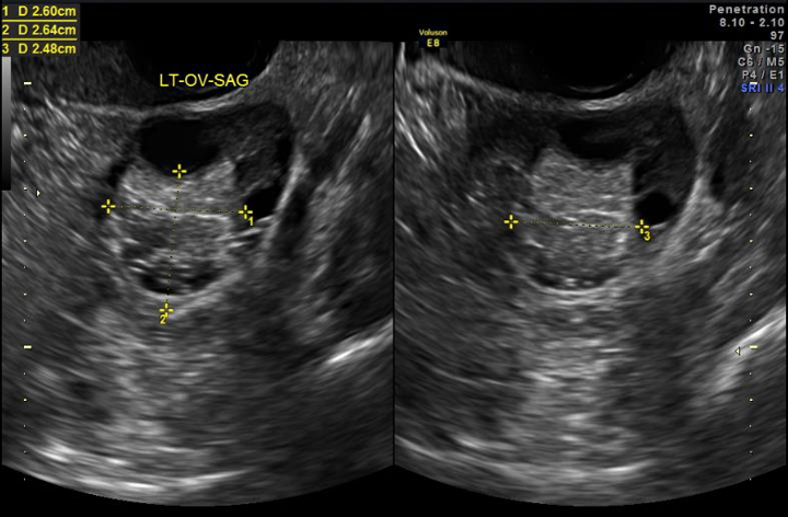
Abnormal ovarian cancer ultrasound images. If cancer is suspected the next step. Imaging tests such as ultrasound or ct scans seen here can help reveal an ovarian mass. A transvaginal ultrasound involves inserting an ultrasound probe slightly larger than a tampon into the vagina to obtain images of the ovaries. 1 2 3 primary ovarian fallopian tube and peritoneal high grade serous ovarian cancer hgsoc high grade serous ovarian cancer is the most prevalent and lethal histologic subtype of.
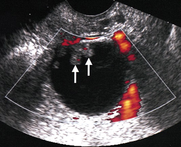
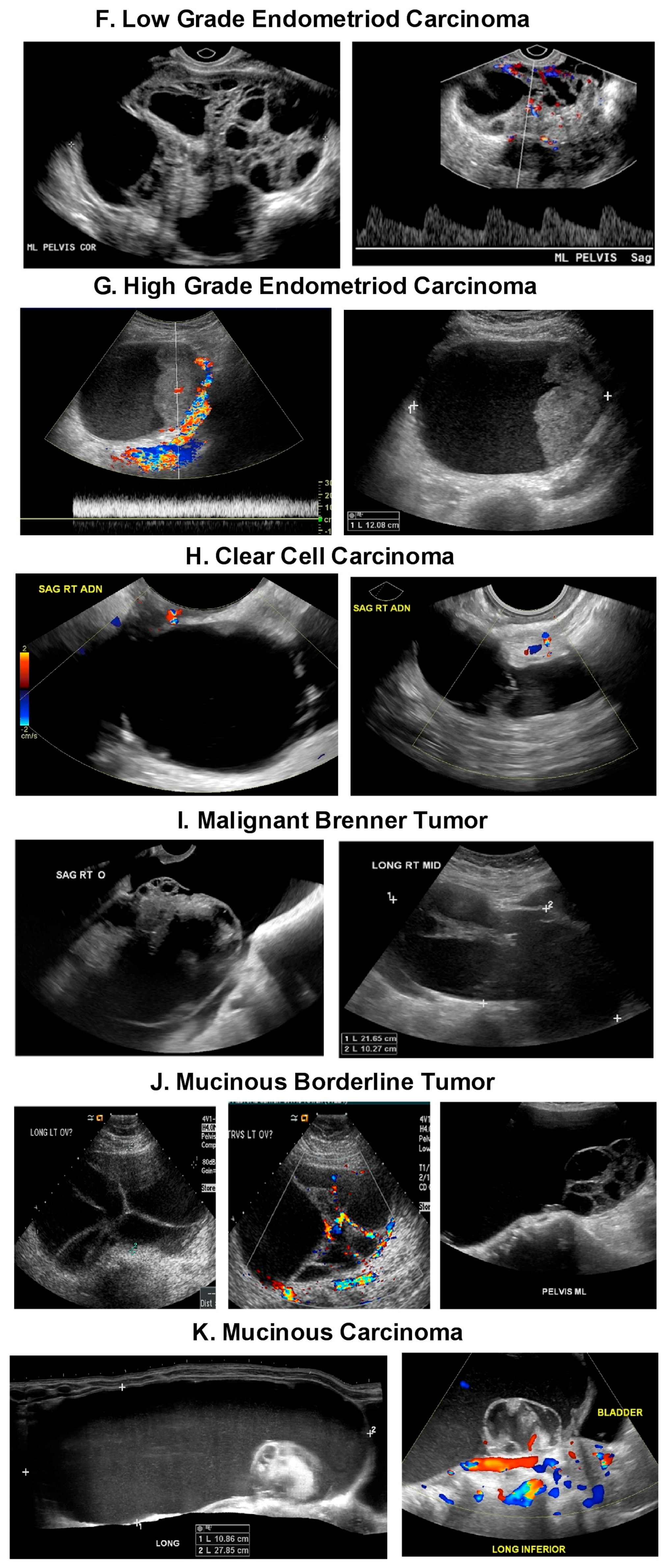
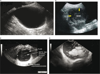
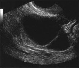
Computed tomography ct scans use x rays to create. The ultrasound appearance of benign and malignant ovarian lesions is shown in figures 1 through 3. 8 11 numerical scores are assigned to morphologic features such as wall structure cyst wall thickness septations and echogenicity. The right ovary in this middle aged female patient see ultrasound images above shows a large primarily cystic mass with multiple septae.
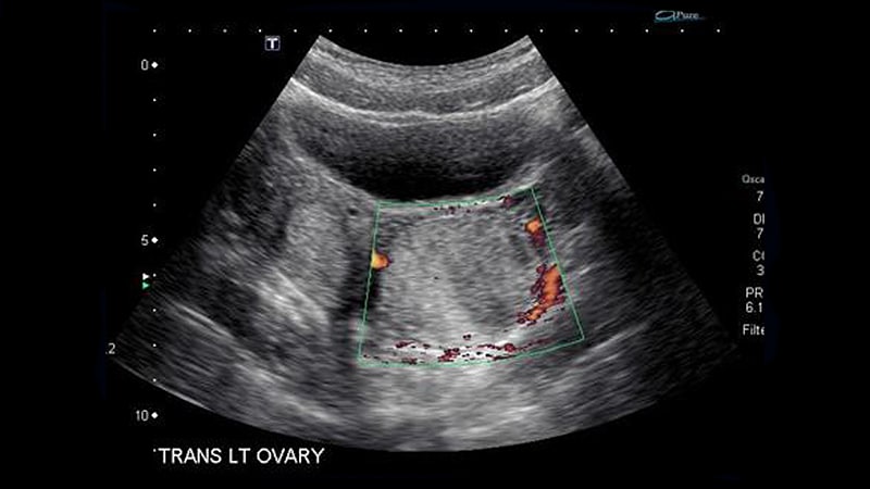
These appearances are typical of mucinous cystadenoma of the right ovary. Researchers are investigating whether transvaginal ultrasound may be effective at screening women who are at high risk of ovarian cancer but who do not have any symptoms of the disease. Some authors have also proposed morphologic scoring systems. However improvements in identification of women at high risk for ovarian cancer as well as improved imaging techniques have increased the likelihood of early detection.
In size and part of it shows coarse particulate matter producing fine echoes within the fluid. It can even see inside the ovaries.
Diagnostics Free Full Text Ultrasound Monitoring Of Extant

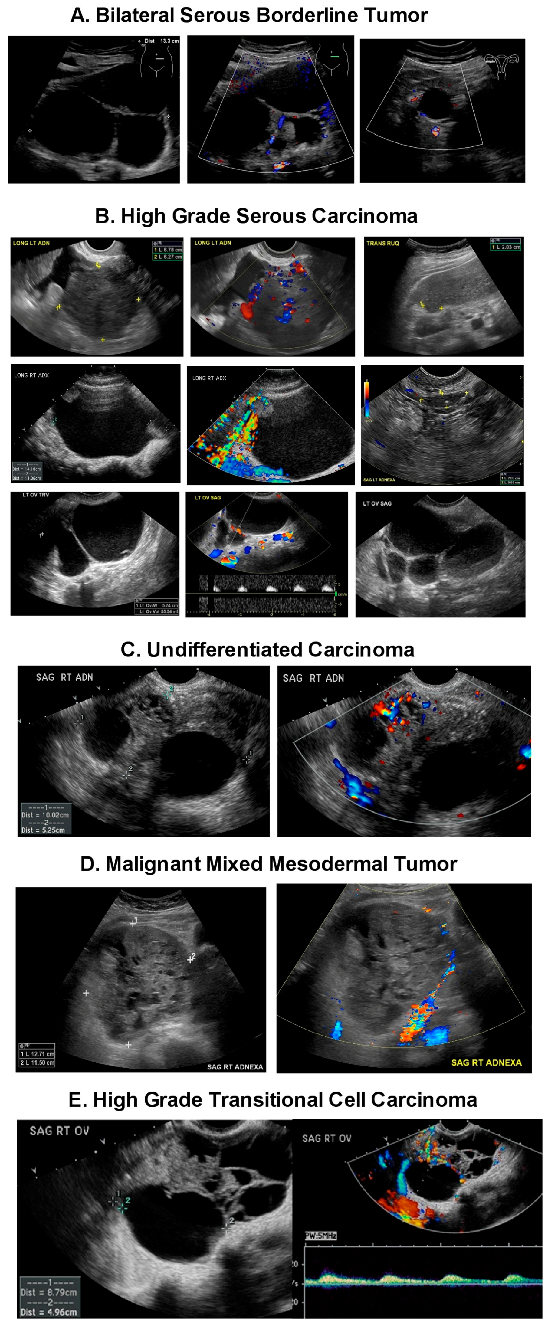
Imaging The Suspected Ovarian Malignancy 14 Cases Mdedge Obgyn
Ultrasound Imaging Of Ovarian Cancer Chapter 22

Imaging The Suspected Ovarian Malignancy 14 Cases Mdedge Obgyn
Detection And Diagnosis Ovarian Cancer Symptoms And Diagnosis

Detection Of Ovarian Tumors In Chicken By Sonography Barua
Plos One Immune Cells In The Normal Ovary And Spontaneous Ovarian

Detection Of Spontaneous Ovarian Tumors At Early Stage With V A
Ovarian Tumors
Normaleah Ovarian Cancer Initiative Detection Diagnosis
Ovarian Cysts
Some Ovarian Tumors Can Be Safely Followed On Ultrasound Cancer
Simple Ovarian Cysts On Ultrasound Need No Further Monitoring